Radiomics uses quantitative pixel information extracted from CT images to assess tumors and enable improved and personalized cancer therapies1. Radiomics research started a decade ago and has rapidly grown since then due to its great potential to predict cancer development and provide tailored treatments2,3. However, inadequate reproducibility of image features in clinical CT imaging impairs the stability and limits the application of radiomics4-6.
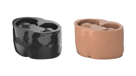
Phantoms are essential for assessing radiomics feature robustness by enabling repeated scanning and comprehensive assessment of quantitative image features in controlled and reproducible settings. Customized phantoms simulating human anatomy and tissue detail were used to evaluate imaging technologies and their impact on radiomics features in reliably characterizing and classifying tissues7,8.
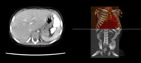
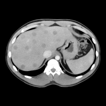
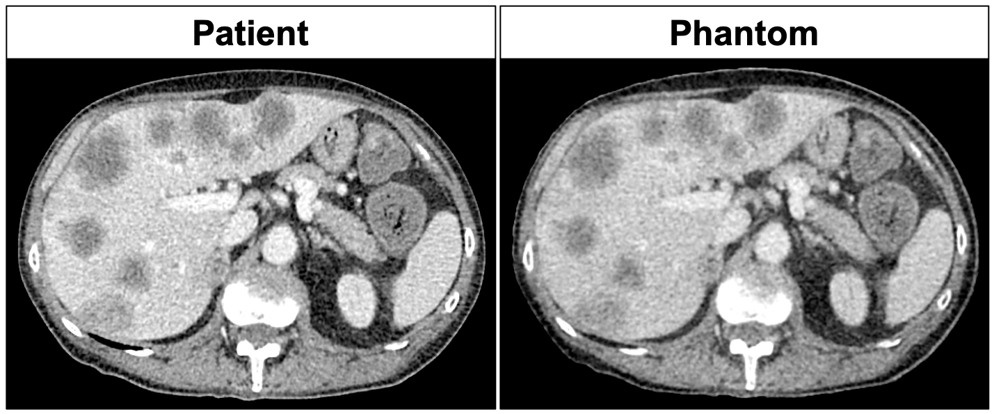
A recently published study investigated the influence of image reconstruction algorithms on radiomics features based on a phantom that reproduced a patient’s abdomen with hepatic metastases9. Study images shown above present the phantom in comparison with the original patient, study images shown below illustrate influences of dose and image reconstruction.
The authors found that filtered back projection and iterative reconstruction algorithms severely compromised radiomics features in accurately and reproducibly characterizing malignant and non-malignant tissues. By contrast, deep learning reconstruction significantly enhanced image quality and yielded a more robust data basis for the extraction of radiomics features. The study highlights limitations of traditional imaging technologies for radiomics, but also shows the potential of incorporating deep learning technologies into image reconstruction to improve image quality for quantitative image analysis.
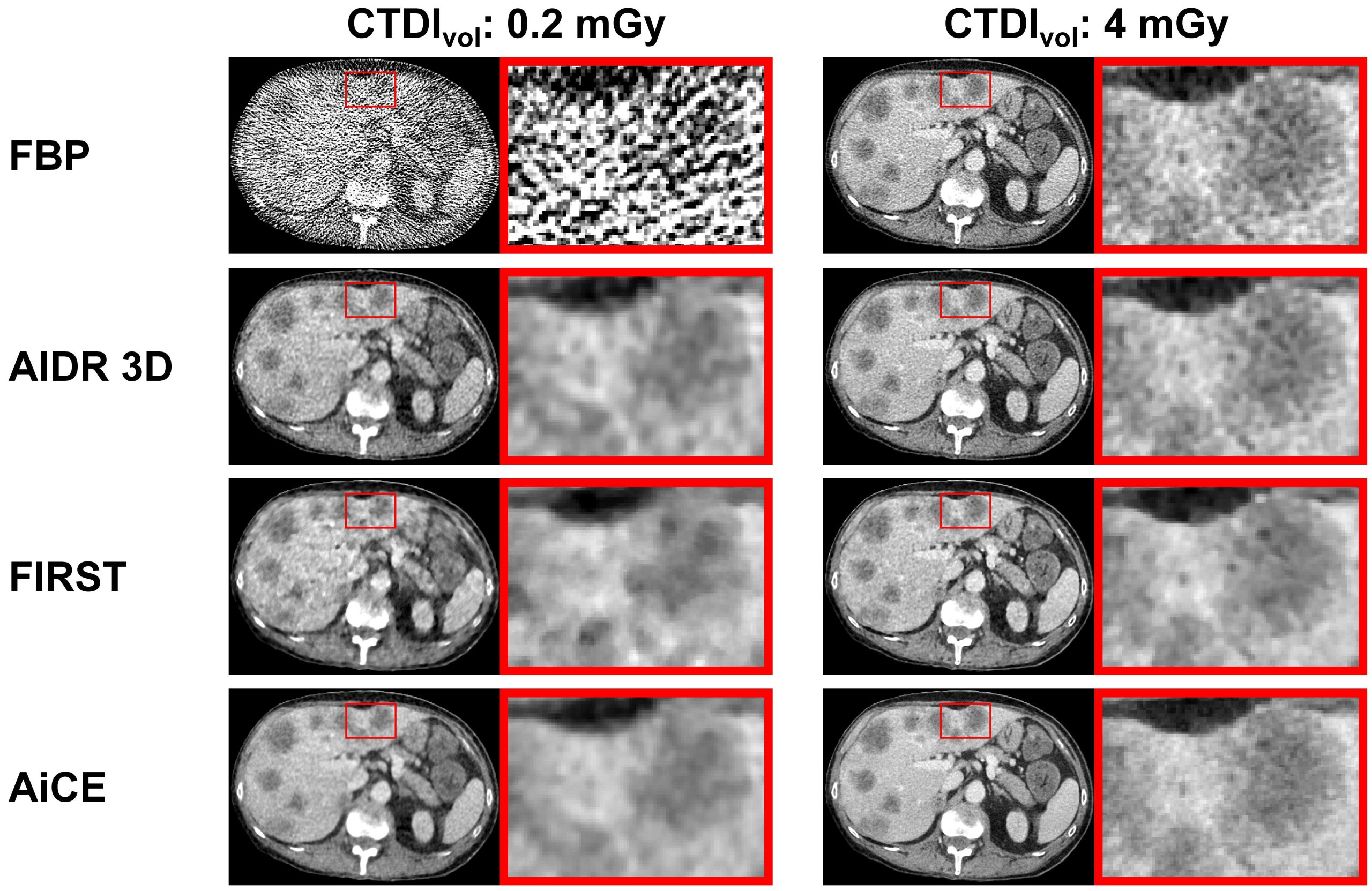
Images originate from Michallek F, et al. Deep learning reconstruction improves radiomics feature stability and discriminative power in abdominal CT imaging: a phantom study. Eur Radiol (2022) and are licensed under Creative Commons Attribution 4.0 International License (http://creativecommons.org/licenses/by/4.0/).
References
- Lambin P, et al. Radiomics: the bridge between medical imaging and personalized medicine. Nat Rev Clin Oncol 14, 749-762 (2017). Link
- Caudell JJ, et al. The future of personalised radiotherapy for head and neck cancer. Lancet Oncol 18, e266-e273 (2017). Link
- Lambin P, et al. Radiomics: extracting more information from medical images using advanced feature analysis. Eur J Cancer 48, 441-446 (2012). Link
- Berenguer R, et al. Radiomics of CT Features May Be Nonreproducible and Redundant: Influence of CT Acquisition Parameters. Radiology 288, 407-415 (2018). Link
- Meyer M, et al. Reproducibility of CT Radiomic Features within the Same Patient: Influence of Radiation Dose and CT Reconstruction Settings. Radiology 293, 583-591 (2019). Link
- Hagiwara A, et al. Variability and Standardization of Quantitative Imaging: Monoparametric to Multiparametric Quantification, Radiomics, and Artificial Intelligence. Invest Radiol 55, 601-616 (2020). Link
- Jimenez-Del-Toro O, et al. The Discriminative Power and Stability of Radiomics Features With Computed Tomography Variations: Task-Based Analysis in an Anthropomorphic 3D-Printed CT Phantom. Invest Radiol, (2021). Link
- Muenzfeld H, et al. Intra-scanner repeatability of quantitative imaging features in a 3D printed semi-anthropomorphic CT phantom. Eur J Radiol 141, 109818 (2021). Link
- Michallek F, et al. Deep learning reconstruction improves radiomics feature stability and discriminative power in abdominal CT imaging: a phantom study. Eur Radiol (2022). Link