Specifications
| Size: | Approx. 234 x 190 x 129 mm |
| Weight: | Approx. 3710 g |
| Base material: | Cellulose-polymer composite |
| Optimal tube voltage: | 120 kVp – adaptable upon request – |


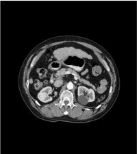
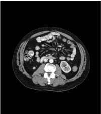
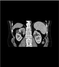
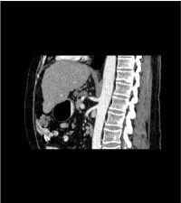
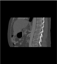

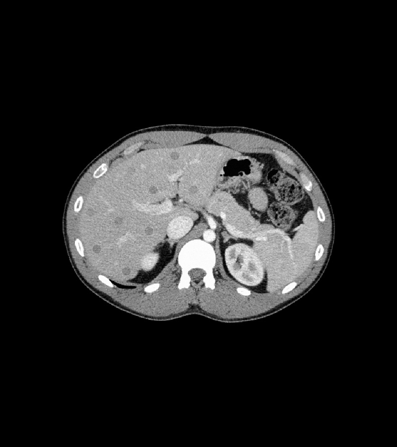
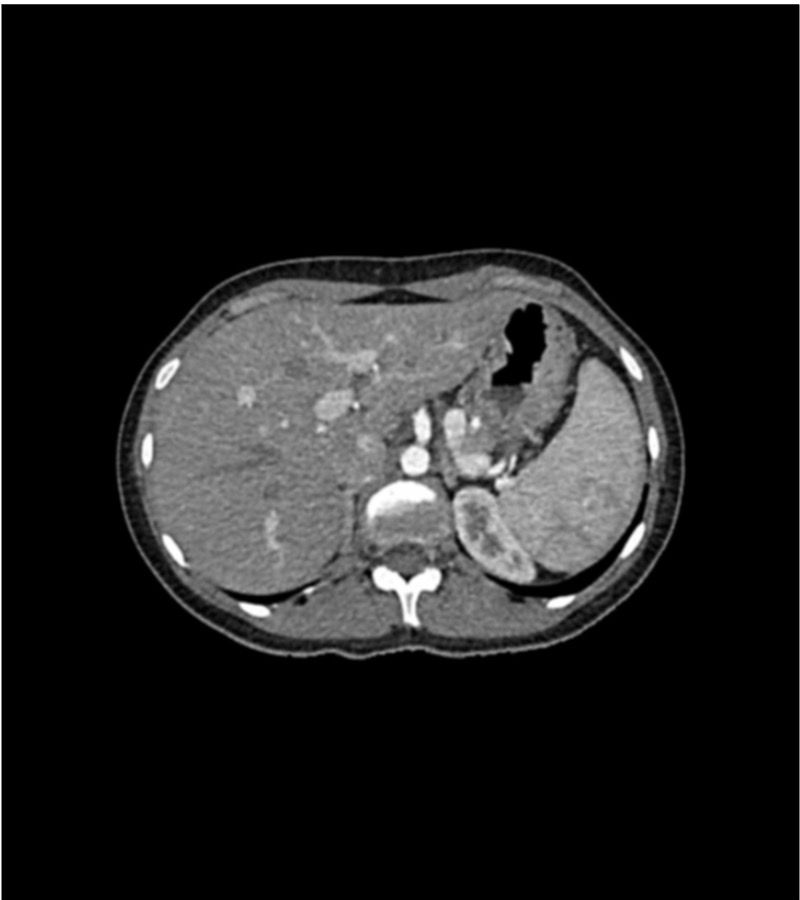
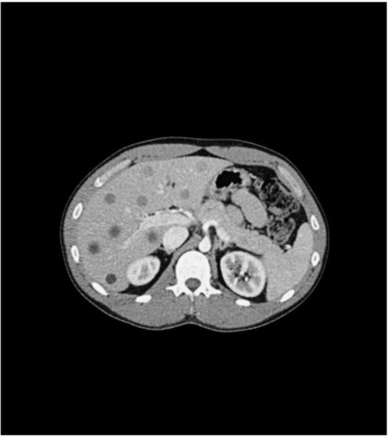
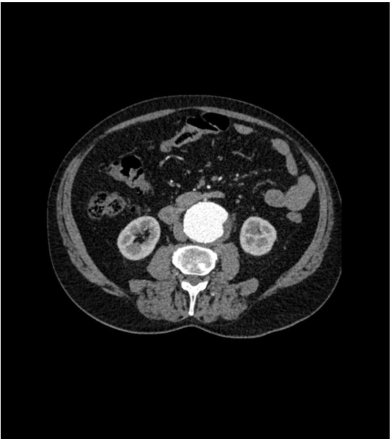
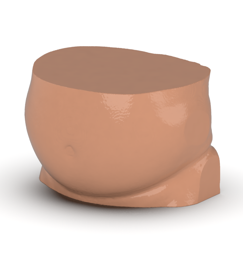
This phantom simulates a contrast medium enhanced abdomen in portal venous phase. It covers the tenth thoracic vertebra to the third lumbar vertebra.
This phantom simulates a contrast medium enhanced abdomen in portal venous phase. It covers the tenth thoracic vertebra to the third lumbar vertebra.
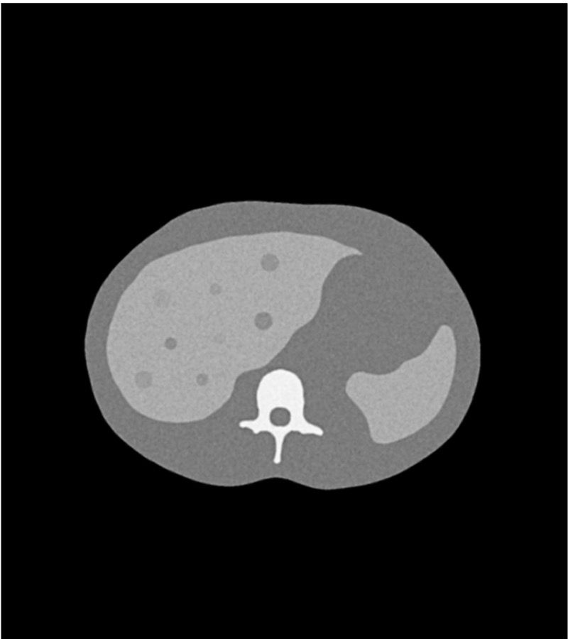
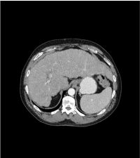
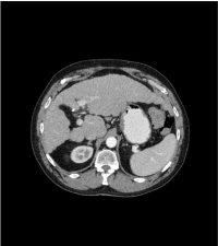
The phantom represents an abdomen after cholecystectomy with small clips. The liver has typical signs of cirrhosis and there is an inferior vena cava filter implanted at the level of the third lumbar vertebra. Both kidneys have cystic lesions and there is a small kidney stone on the left side.
The phantom can be used in CT (including CBCT) to evaluate and optimize imaging performance and post-processing applications, including AI-enabled applications. It is also suited for training purposes.
The phantom provides a detailed and realistic simulation of soft and bone tissue. Air voids are filled with a cellulose-polymer composite of approx. -160 HU.
| Size: | Approx. 234 x 190 x 129 mm |
| Weight: | Approx. 3710 g |
| Base material: | Cellulose-polymer composite |
| Optimal tube voltage: | 120 kVp – adaptable upon request – |
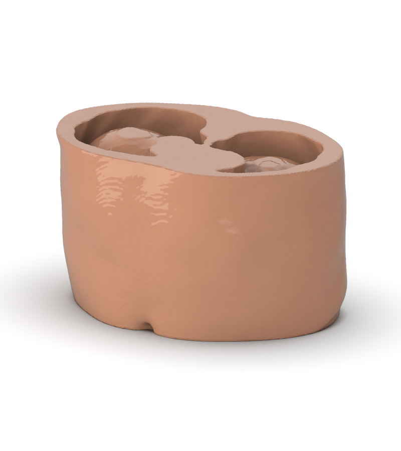
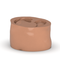
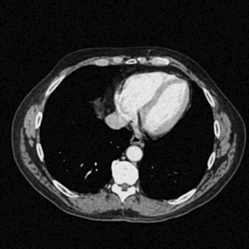
Realistic simulation of vasculature, bone and soft tissues, including the liver, pancreas, spleen, adrenals, kidneys, stomach, small intestine and colon.
Liver cirrhosis, cholecystectomy, inferior vena cava filter, kidney cysts, kidney stone, lymph nodes.
The product ships in a protective hard case featuring interior foam padding.
Dimensions (case): 415 x 280 x 190 mm
Total weight (ready to ship): 10.0 kg
*Outer packaging (e.g., cardboard box or protective wrap) may vary slightly, affecting total dimensions.