Head Phantom CTA AVM Lesion

Data 34 doses

SKU: D50-05-1
Scanner: Canon Aquilion ONE
Size (GB): 62,31
Number of images: 126.694
Number of protocols: 238

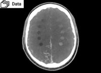
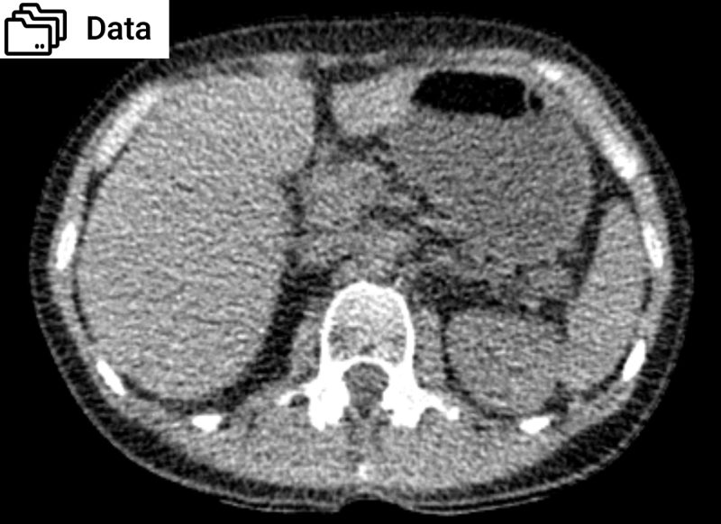
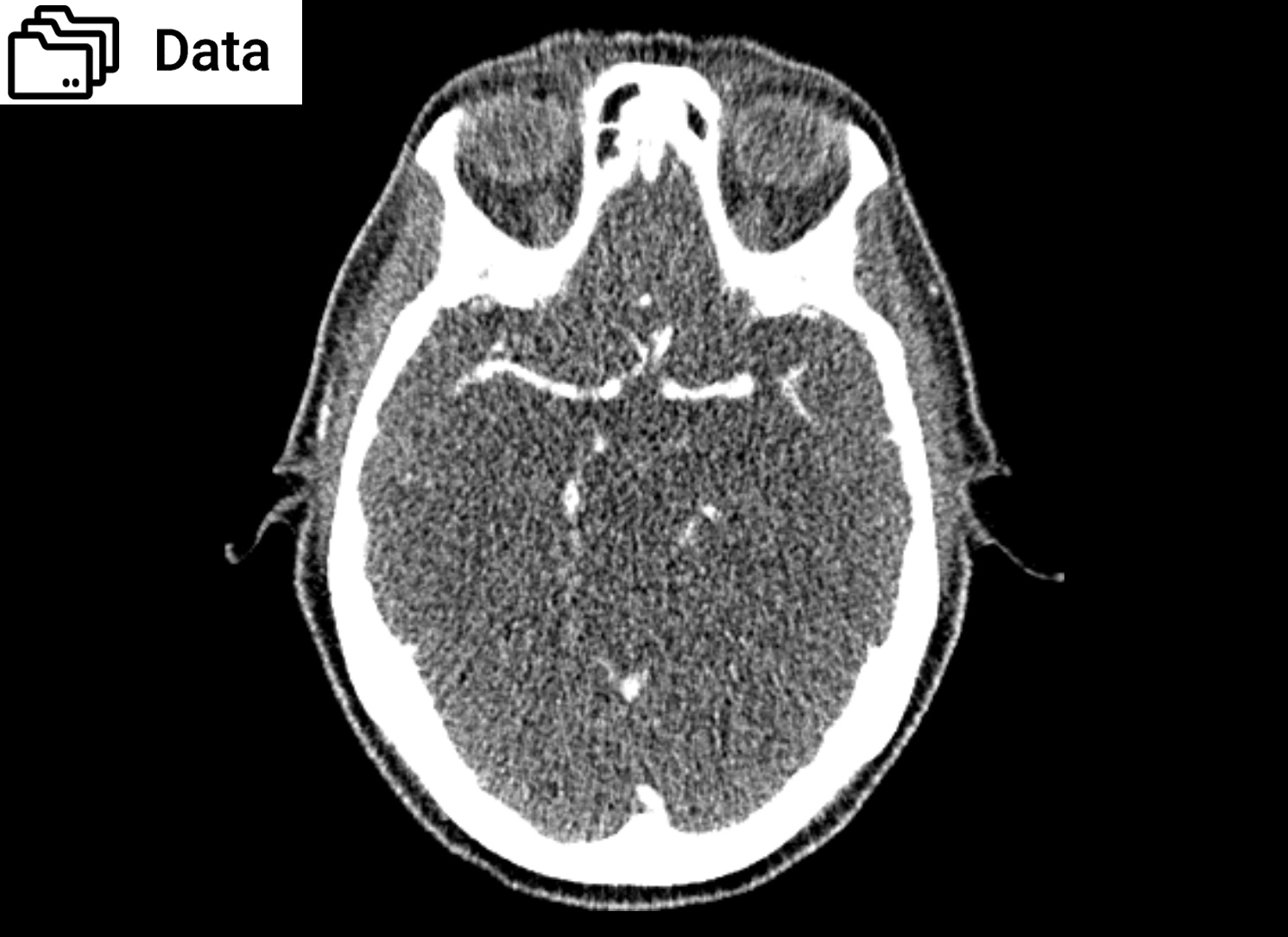
This data set comprises 126.694 axial images of head CTA phantom AVM / lesion 50-05 acquired with a Canon Aquilion ONE scanner.

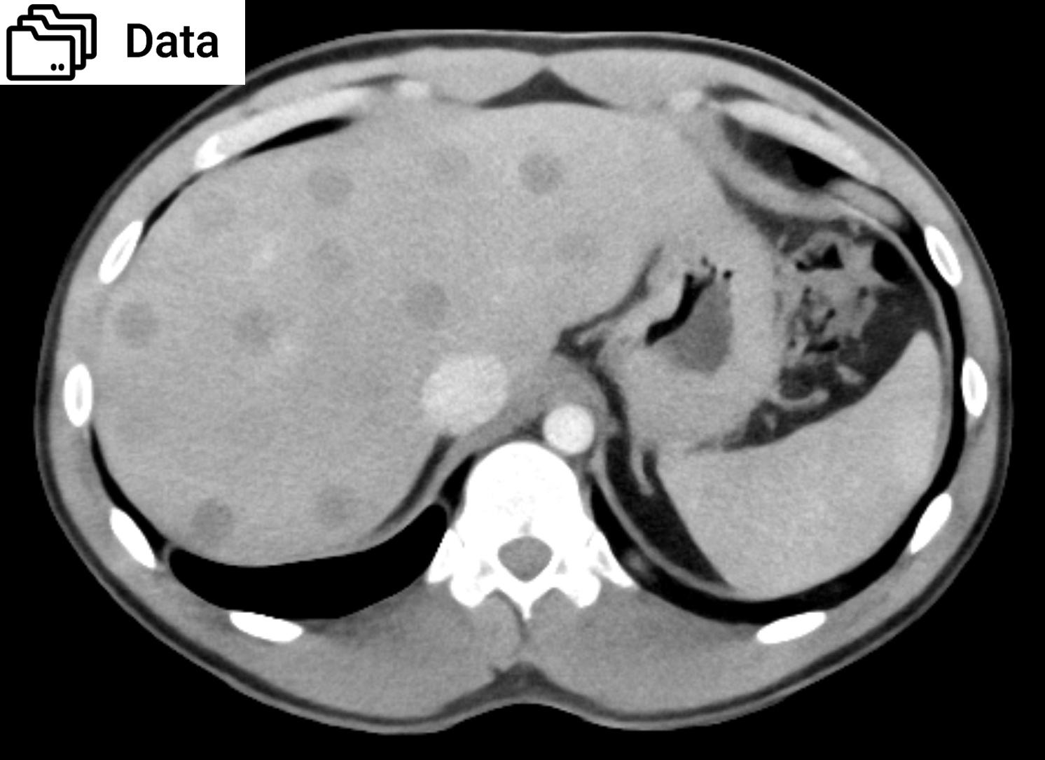
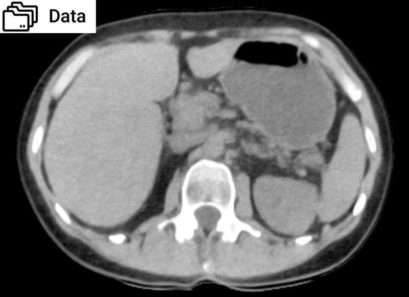
The data is organized in 238 series with 34 doses from 0.3 to 25.5 mGy and five reconstruction methods: Filtered back projection (FBP FC26), standard hybrid iterative reconstruction (AIDR 3D FC26), model-based iterative reconstruction (FIRST), and deep-learning reconstruction (AiCE). Two repeated acquisitions of each series are included. AIDR 3D, FBP and AiCE acquisitions are available with a slice thickness of 1mm and 0.5mm, FIRST only in 0.5mm.

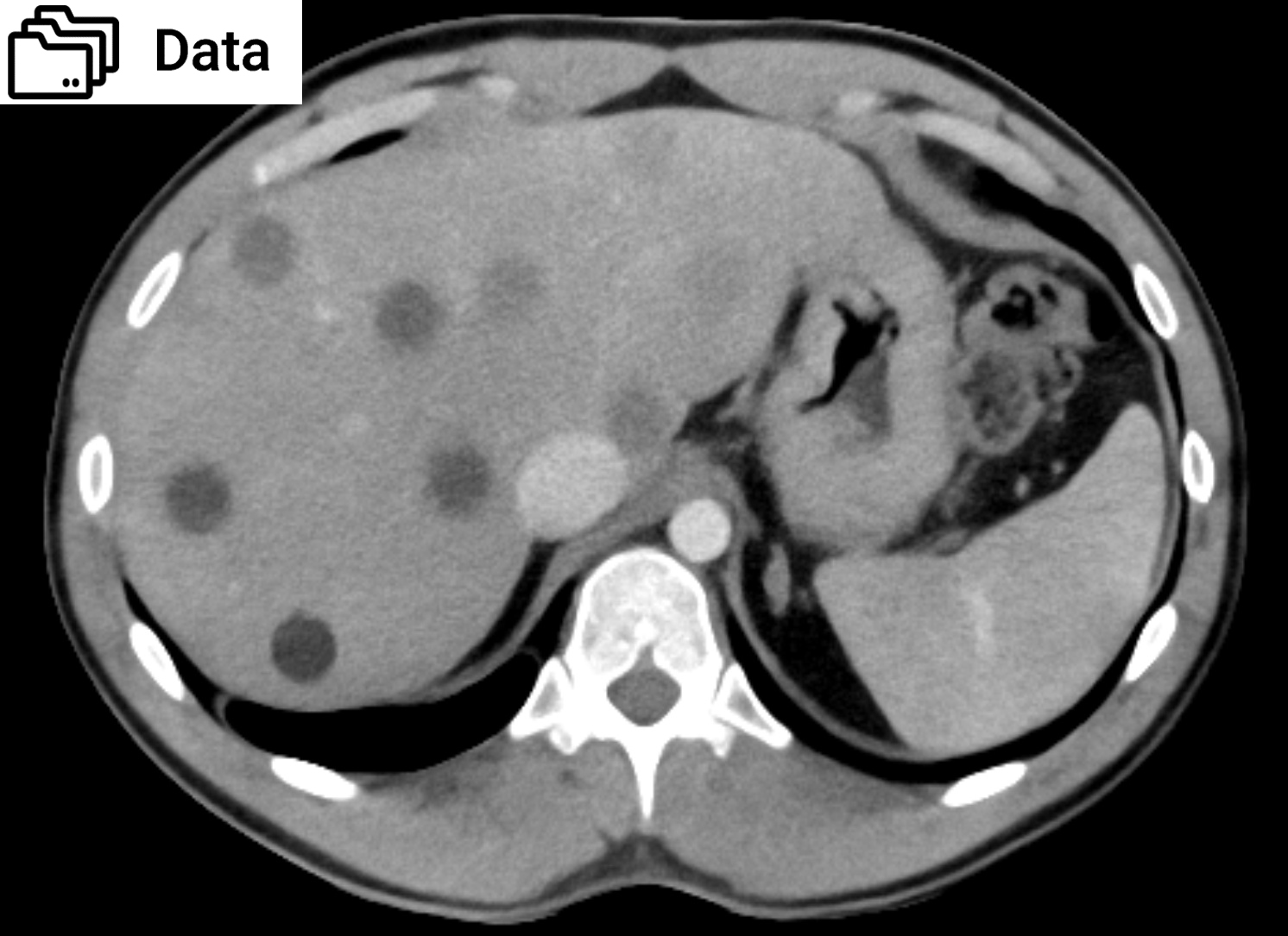
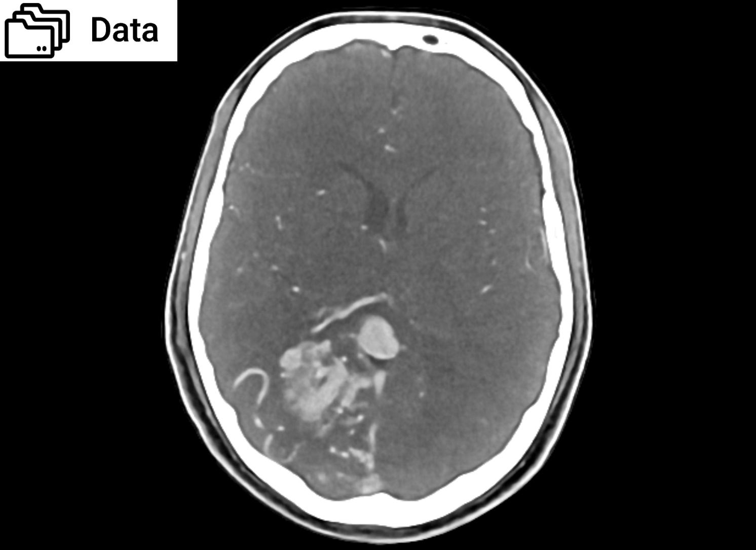
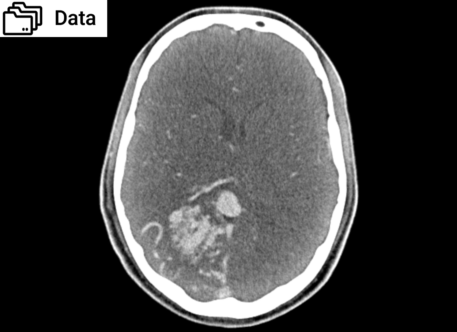
The data provides comprehensive coverage of anatomy and pathology (arteriovenous malformation of the right hemisphere, low-contrast lesions) across various reconstruction methods in state-of-the-art CT imaging.


Specifications

Scanner: Canon Aquilion ONE
Size (GB): 62.31
Number of images: 126694
Number of series: 238
Repetitions per series: 2
Images per series: 163  (1mm) / 325 (05mm)
Image plane: Axial
Tube voltage (kVp): 120


CTDI (mGy): 0.3, 0.7, 1.0, 1.4, 1.7, 2.1, 2.4, 2.8, 3.1, 3.5, 3.8, 4.2, 4.5, 4.9, 5.2, 5.6, 5.9, 6.3, 6.6, 7.0, 7.3, 7.7, 8.0, 8.4, 8.7, 9.1, 9.4, 9.8, 10.1, 10.5, 14.6, 18.2, 21.9, 25.5
Tube current (mA): 10, 20, 30, 40, 50, 60, 70, 80, 90, 100, 110, 120, 130, 140, 150, 160, 170, 180, 190, 200, 210, 220, 230, 240, 250, 260, 270, 280, 290, 300, 400, 500, 600, 700
Rotation time (s): 0.5
Slice thickness (mm): 0.5, 1
Slice increment (mm): 0.5, 1
Kernel types: Soft tissue, Bone
Image reconstruction: FBP, AIDR 3D, AiCE, FIRST


Data Sheet

DOWNLOAD DATA SHEET