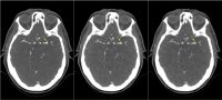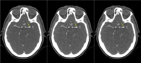
Iterative reconstruction was introduced into clinical routine more than a decade ago and is a standard reconstruction method widely used in today’s CT imaging. Iterative reconstruction methods have strong denoising power and can maintain reasonable noise levels in images acquired with reduced doses. Studies have reported significant dose reduction potential with iterative reconstruction compared to images reconstructed with conventional filtered back projection. However, image noise and subjectively perceived image quality are limited predictors of diagnostic image quality, when iterative reconstruction is used1. In particular, low image noise can suggest high image quality although findings are not accurately displayed2.
Task-based methods of image quality assessment test whether CT images present findings with sufficient accuracy to enable diagnostic tasks (e.g., lesion detection, segmentation, classification) and have been recommended for objectively assessing CT imaging technologies, particularly when using modern denoising reconstruction algorithms that have nonlinear properties such as iterative reconstruction1. Task-based methods have been reported to provide the best prediction of clinical imaging performance3, with realistic phantoms and test scenarios being relevant to ensure transferability of CT assessment to clinical imaging3-5.
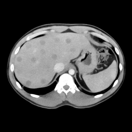
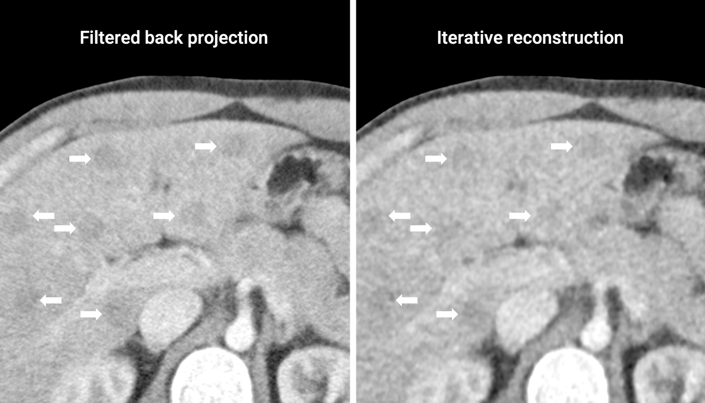
The two images shown above have equivalent contast-to-noise ratios of liver lesions and liver parenchyma, but illustrate differences in hepatic lesion conspicuity and delineation despite powerful denoising of iterative reconstruction. Both images show the same abdomen phantom (based on phantom 508-02) with low-contrast liver lesions of 11 mm diameter, imaged with a CTDIvol of 7 mGy (left) and a CTDIvol of 1 mGy (right).
References
- Vaishnav JY, et al. Objective assessment of image quality and dose reduction in CT iterative reconstruction. Med Phys 41, 071904 (2014). Link
- Mileto A, et al. State of the Art in Abdominal CT: The Limits of Iterative Reconstruction Algorithms. Radiology 293, 491-503 (2019). Link
- Samei E, et al. Performance evaluation of computed tomography systems: Summary of AAPM Task Group 233. Med Phys 46, e735-e756 (2019). Link
- Solomon J, et al. Comparison of low-contrast detectability between two CT reconstruction algorithms using voxel-based 3D printed textured phantoms. Med Phys 43, 6497 (2016). Link
- Conzelmann J, et al. Comparison of low-contrast detectability between uniform and anatomically realistic phantoms-influences on CT image quality assessment. Eur Radiol 32, 1267-1275 (2022). Link